บทความ
Review article: Mucous membrane pemphigoid Part II; Differential diagnosis and management
Differential diagnosis
Clinical presentation of mucous membrane pemphigoid (MMP) should be differential diagnosis from the other vesiculobullous diseases such as pemphigus vulgaris (PV), bullous pemphigoid (BP), epidermolysis bullosa (EBA), linear IgA disease and others mucocutaneous diseases such as lichen planus, bullous systemic lupus erythematosus and erythema multiforme. MMP and BP have the same histopathologic features and direct immunofluorescence (DIF), so the differential diagnosis should be made on the combination of clinical findings and indirect immunofluorescence (IIF) examination. MMP predominantly involves the mucous membrane, whereas BP typically affects the skin. In addition, circulating antibodies in BP are more common than in MMP [1].
EBA and MMP may also present the same clinical, histopathological and immunopathological features. The distinction can be achieved by salt-split skin technique using human skin. If antibodies deposit on the roof side of the induced separation, the diagnosis is most possibly to be MMP. In contrast, if the deposition is on the floor (dermal side), the diagnosis is EBA [2].
The general features of MMP compared with PV and BP are showed in table 1.
Table 1. General features of PV, MMP and BP (Modified from Xu et al., 2013 [1] and Pongsiriwet et al., 2018 [3])


MMP Management
Treatment of MMP is based on the involved sites, severity and disease progression. Additionally, it should be individualized depending on age, medical history and contraindications of any systemic medications [1]. Early diagnosis and treatment may decrease disease-related complications, especially airway obstruction, stricture and blindness [4]. In low-risk patients with lesions affecting oral mucosa and/or skin can be treated effectively with topical therapy, such as topical corticosteroids or topical calcineurin inhibitors. For more severe or recalcitrant lesions or during exacerbation of disease in low-risk patients, the treatment should be combined with systemic therapy. High-risk patients with rapid progression or multiple involving sites including ocular, genital, esophageal or nasopharyngeal mucosa require more aggressive systemic treatment with topical treatment. A multidisciplinary approach including oral medicine experts, ophthalmologists, gastroenterologist, otolaryngologist, gynaecologist, and dermatologists is essential for the management of MMP and related to the treatment outcome [4].
Low-risk patients
Potent topical corticosteroids are advised initially, applied 2-3 times/day. In patients with isolated severe or recalcitrant lesions, intralesional corticosteroid injections with triamcinolone acetonide 10mg/ml 0.1 cc/cm2 can be used. For desquamative gingival lesions, topical corticosteroids in gel form are recommended and should be applied with custom tray which covers the involved gingiva [1]. Additionally, combination therapy of topical tacrolimus with prednisolone 40mg/day has been shown to be effective after 3 months of treatment [5]. If the patients do not response to topical therapy, dapsone (50-200 mg/day) or tetracycline (1-2 g/day) or nicotinamide (2-3 g/day) can be added. For non-responsive patients with above regimens, systemic corticosteroids such as prednisolone (1-2 mg/kg) can be used [6]. Systemic corticosteroids may be combined with immunosuppressive agents such as azathioprine (1-2 mg/kg/day) or mycophenolate mofetil (1-2 g/day) [7].
High-risk patients
Systemic corticosteroids in combination with immunosuppressive drugs are the treatment of choice for severe or rapidly progressive disease [4]. Prednisolone 1-1.5 mg/kg/day combined with cyclophosphamide 0.5-2 mg/kg/day is recommended. Alternatively, prednisolone can be added with other immunosuppressive agents such as mycophenolic acid 2-2.5 g/day or azathioprine 1-2 mg/kg/day. For mild disease, dapsone 50-200 mg/day can be given [1]. When the disease becomes effectively controlled, prednisone should be tapered gradually while continuing immunosuppressive drug. Generally, the adjuvant immunosuppression should be continued for 2 years [6]. In patients with progressive ocular lesions refractory to corticosteroids and immunosuppressive drug, intravenous immunoglobulins can be used to prevent complications, especially blindness [8]. Additionally, biologic agents such as rituximab, etanercept or TNF-alpha inhibitors have been reported to treat severe and recalcitrant MMP successfully and should be an alternative treatment option [6]. Long-term treatment with prednisolone can cause several side effects, so carefully monitoring should be performed appropriately [4].

CR; complete response, PR; partial response, NR; no response)
Figure 1. Treatment algorithm for MMP
(Modified from Xu et al, 2013 [1], Pongsiriwet et al, 2018 [3], and Bagan et al [9])
Conclusion
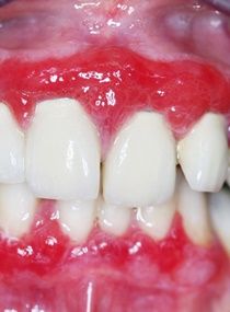
MMP is a chronic autoimmune disease characterized by subepithelial blisters that typically affects mucous membranes more often than skin. It is more common in female and mainly affects elderly people. The most frequently involving site is the oral cavity, followed by conjunctiva. Clinical presentation and severity of MMP patients are highly variable. Clinical manifestations in the oral cavity include desquamative gingivitis, blisters, erythema, erosions and ulcerations. Diagnosis is mainly based on clinical findings, histopathologic examination and immunofluorescence studies. There is no gold standard therapy for MMP. The treatment depends on the sites of involvement, clinical severity and disease progression. Topical therapy is the mainstay of treatment for localized disease. For more severe and widespread disease, more aggressive and systemic therapies are the treatment of choice. Scarring is commonly seen, especially in oropharyngeal and ocular mucosa which can progress to esophageal and laryngeal stenosis, strictures and blindness. Early diagnosis and treatment may decrease disease-related morbidity and mortality. Multidisciplinary approach is necessary for the diagnosis and management of MMP.