บทความ
Surface Electromyographic Studies on Masticatory Muscle Activity
Related to Orthodontics: A Review of Literature
Abstract
The understanding of masticatory muscle function and its relationship with craniofacial morphology is essential to the field of orthodontics. Electromyography (EMG) has been used to assess muscle function both qualitatively and quantitatively. Many studies attempted to relate masticatory muscle activity with facial form, but the results have been inconsistent. The influence of the masticatory muscles on orthodontic treatment, specifically vertical malocclusion correction, and stability is still controversial. The purpose of this article is to review the relationship among masticatory muscle function, facial morphology and malocclusion based on the electromyographic studies.
Introduction
The form and function of masticatory muscles are believed to correlate with the craniofacial growth and subsequent orthodontic and dentofacial orthopedic treatments.1-4 Awareness of skeletal muscular environment is necessary because orthodontic treatment plan is not dependent exclusively on biomechanical factor. Masticatory muscles could affect the active treatment of malocclusions and jaw deformities, as well as the stability of such treatment.5
With relevance to orthodontics, the main muscles associated with mastication are anterior and posterior temporal, superficial and deep masseter, superior and inferior lateral pterygoid, medial pterygoid and digastric muscles. Surface electromyography (EMG) is a reliable non-invasive tech¬nique for evaluating muscle activity by detecting the electrical potentials via electrodes placed over the skin. Surface EMG has gained popularity in dentistry due to the ease of the procedure.6 However, the use of surface EMG in examining superficial muscle action depends on both individual’s physiology and recoding techniques.6
EMG studies seeking to link masticatory muscle activity with facial form, have given various results4,7,8 and it is difficult to accurately define such association because of large inter-individual variability and many etiological factors for malocclusions. Vertical malocclusion could result from the interaction of different etiological factors. Masticatory muscle activity may be one factor affecting the bite opening in deep bite correction. There is still much controversy regarding the complex relationship between masticatory muscle features and vertical facial pattern.
This article presents the use of surface electromyography in studying the association between masticatory muscle and craniofacial morphology. Moreover, changes in muscle activity following orthodontic treatment are also discussed.
Basics of electromyography
In assessment of muscle function, one of the most common recording techniques is electromyography (EMG). The record obtained from electromyography is called Electromyogram9 (Fig. 1). This technique assesses the magnitude and duration of muscle activity by recording the intrinsic electrical potential arising from the active motor units.6 It has been used to evaluate muscle function both qualitatively and quantitatively.10, 11 During both static and dynamic conditions, researchers have gained knowledge of how individual muscles are controlled.

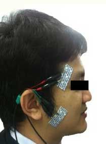
There are two main types of EMG (Fig.2). The first one is intramuscular electromyography in which needles or fine-wire electrodes are inserted through the skin into the muscle. These electrodes can record single motor unit action potentials. The other type is surface electromyography (sEMG), which uses surface electrodes to detect superimposed and summated motor unit action potentials. Surface EMG is technique-sensitive. It’s the accuracy and reliability depends on the position of the electrodes, skin impedance and external noise. The surface electrodes may spontaneously record the signals from nearby muscles. However, sEMG from the superficial masseter muscle and the anterior temporal muscle, has shown acceptable results.12

Both needle and surface EMG have their pros and cons. Needle electrodes have less technical artifacts than surface electrodes since the distance between the electrode and the muscle is constant. However, the needle electrodes may cause infection and discomfort. Moreover, the activity of the whole muscle cannot be recorded. Surface electrodes, on the other hand, are less invasive and reduce the risk of infection. The signals obtained from the surface electrodes are considered to represent the activity of the whole muscle or muscle groups. Since electrode placement can be visually monitored, surface recordings are believed to be more reproducible than those obtained intramuscularly.10 Disadvantages of sEMG are non-specificity resulting from signals recorded from adjacent muscles and increased general noise level, and the loss of signals due to skin properties.13 Additionally, the errors may increase when the distance between the electrodes and the muscle is not constant during muscle contraction.14 It is not possible to compare amplitudes of raw EMG signals. Thus, it is problematic to make quantitative comparisons.11
Factors affecting EMG recordings
Normalization of the raw EMG signal to the maximum voluntary contraction level could be used when the comparison is needed. Surface EMG has been used widely in physiological investigations of the masticatory muscles.14 However, quantification of surface EMG recording is dependent on many factors; for example, individual variation in muscle fiber depolarizations, electrode placement location relative to the muscle being studied, skin-electrode contact quality, and signal diminishing characteristics of the intervening tissues.13 Changes in the placement of surface EMG electrodes or preparation of the skin to improve contact and reduce skin impedance by scrubbing the skin and wiping it with alcohol can increase the magnitude of the signal recorded.14 EMG signal processing includes filtering of background noise, modification, smoothing or averaging and integration. However, signal processing does not certainly prove the quantitative comparison of EMG results. Because the magnitude of the recorded signal depends on the distance from the generated signal and on the characteristics of the intervening tissues, amplitudes difference in the same muscle in different individuals and between muscles of the same individual.
Tabe et al. compared the activity of masticatory muscles assessed during the day and at night. They found a decrease in the EMG activity at night.15 Garnick examined three normal subjects under identical conditions at 20-minute intervals and again after two weeks.16 The differences of surface recordings from the masseter muscle during mastication were related to the time between recordings. Moreover, Ueda et al. recorded sEMG for 24 hours and demonstrated a longer duration of the activity of the temporalis muscles in children and the masse¬ter muscles in adults.17 This might be because of incomplete development of the dentition and the immaturity of the muscles in children. Ferrario et al. showed that the EMG activity of the anterior temporalis and masseter muscles in healthy males and females were similar during rest position and centric occlusion, but different during maximum voluntary clenching (MVC) where males had higher EMG activity than females.18 Moreover, the masseter muscle had more asymmetrical EMG activity than the temporalis muscle. In this study, the masseter activity was stronger in clenching and dominant for the temporalis activity in centric occlusion and rest position in males. In contrast, the temporalis muscle activity in females tended to dominant at every contraction level.
Many investigators have examined muscles at rest, during mastication, and during swallowing.10,11,19 However, mastication and swallowing are complex functions that are difficult to control. The mandibular movements may vary in magnitude and speed from subjects to subjects, or even within the same subject at different times. The level of jaw opening and velocity of contraction have affected recorded EMG magnitudes and patterns.20 The studies of EMG in orthodontics usually record EMG of the masticatory muscles before, during, and after the application of functional orthodontic appliance so as to monitor or assess their effectiveness. The activity of the temporalis and masseter muscles during clenching, chewing, and swallowing have been studied.
Table 1 Summary of factors affecting the EMG signal|
Technical factors |
|
|
Physiological factors |
|
Masticatory muscle activity and malocclusion
Moyers was the first one who attempted to apply electromyography to dentistry.12 He observed that muscular balance influenced the normal relation of teeth. In orthodontics, the temporalis and masseter are two major superficial muscles that were used to represent masticatory muscle function in sEMG studies. Christensen et al. revealed that during maximal clenching, the masseter muscle provides most of the isometric force, while the posterior temporalis is the main postural muscle, irrespective of the type of malocclusion.21
Masticatory muscle activity and malocclusion in the vertical dimension
The association of vertical malocclusions on the electrical activity has been studied. Ciccone de Faria et al. compared the EMG activity of masticatory muscles (the temporal and masseter muscles) in skeletal or dentoalveolar open bite children, aged 6–11 years, with the control group.22 EMG was performed under maximal voluntary clenching and during chewing. They found that the control group presented the highest EMG signals in the temporalis and masseter muscles during MVC (85.2 %). Subjects with a dentoalveolar anterior open bite demonstrated significantly lower temporalis and masseter muscle percentage activity (61.52 %). Patients with a skeletal open bite malocclusion showed the lowest electrical activity (42.13 %), especially during chewing.
Similarly, the study by Yousefzadeh et al. recorded EMG activity of the temporalis, masseter, orbicularis oris and digastric muscles in patients with an anterior open bite, aged 10–13 years.23 During clenching, patients with an anterior open bite presented lower EMG activity on the working side compared with the controls. During chewing, the anterior open bite group tends to show higher EMG activity on the balancing side of the temporalis and masseter when compared with the normal subjects. It is also possible that the functional efficiency of the muscles in anterior open bite patients is lower and lacks canine and incisal guidance, or the balancing side interferences activate more of balancing side muscles during mastication. Thus, more or larger motor units are shown from both sides to generate the necessary force during mastication. Likewise, Ciccone de Faria et al. showed that the control and the dentoalveolar anterior open bite groups presented a higher EMG activity when clenching, which differed from the mean values when chewing, but no difference in EMG activity between clenching and chewing in the skeletal open bite group was found.22 Based on these studies, it may be suggested that the maximal activity of masticatory muscles is decreased with anterior open bite. Reduced occlusal stability, increased vertical dimension and weak masticatory muscles were found in subjects with an anterior open bite.
Ahlgren showed the EMG activity during chewing in 80 children, but found no significant correlations between cephalometric measurements and integrated EMG activity. However, there was a tendency to a negative correlation (r = -0.3) between EMG activity and the gonial angle.24 Ahlgren et al. also studied the maximum voltage amplitudes of surface EMG recordings from the masseter, anterior, and posterior temporalis and showed that patients with acute gonial angles had lower levels of masseter muscle activity during swallowing, and higher anterior temporalis activity at rest than those with obtuse gonial angles.25 Similarly, the significant association was found in the study of Lowe and Takada in that a higher resting level for masseter activity was observed in subjects with short mandibles and steep occlusal planes.
Masticatory muscle activity and malocclusion in the sagittal dimension
Table 2 The association of sagittal malocclusions in different Angle molar classification on the electrical activity was studied|
Author |
Study Groups |
Sample |
Age, y |
Methods |
Main findings |
|
Moreno et al. 26 |
I: right Class I II: right Class II III: right Class III IV: left Class I V: left Class II VI: left Class III
|
12 males, 33 females n = 31 n = 8 n = 6 n = 30 n = 8 n = 7 |
(22-29) 24 |
Raw EMG activity of the masseter, anterior and posterior temporalis, digastric muscles during MVC, swallowing and chewing
|
- The values of temporalis EMG activity in MVC for patients with Angle molar Class I, II, and III were significantly different: 185.40 μV, 123.46 μV, and 226.80 μV, respectively. - Patients with Class II showed higher EMG activity of the temporalis muscles than other classes in deglutition and chewing. - Subjects with Class III achieved the highest activity for the temporalis and masseter muscles during MVC. |
|
Cha et al. 27 |
I: Class I malocclusion, normodivergent II: Class I, hyperdivergent III: Class II, normodivergent IV: Class II, hyperdivergent V: Class III, normodivergent VI: Class III, hyperdivergent |
38 males, 67 females n = 27 n = 20 n = 10 n = 23 n = 12 n = 13 |
(22.0 + 6.7) |
Raw EMG activity of the masseter muscles and anterior temporalis during resting and clenching
|
- No significant differences of masseter activity in resting among all groups except resting for anterior temporalis activity was significantly higher in patients with Class III and SN-GoMe>36°. |
|
Ahlgre-n et al. 25 |
I: Class II division 1 II: Normal occlusion |
Males n = 15 n = 15 |
(9-11) 10.6 (9-13) 10.11 |
Raw EMG activity of the masseter, orbicularis oris, anterior and posterior temporalis muscles during resting, chewing and swallowing |
- No difference between types of occlusion in the EMG activity was found in the rest position, which was greatest for the posterior temporalis muscle for both types of occlusion. - Class II occlusion group had a tendency to develop less EMG activity during chewing than normal occlusion group. - Posterior temporalis activity dominated during rest, and anterior temporalis during mastication. - Less EMG activity in the anterior temporalis and masseter was found during swallowing compared with the normal group |
|
Moyers 12 |
I: Class II division 1 II: Normal occlusion |
n =16 n =16 |
(4.6-16) |
Raw EMG activity of the temporalis, masseter, internal and external pterygoid, suprahyoid, mental muscle during resting, occluding and moving |
- Variation of the spike potentials of EMG was found, particularly when the temporalis muscle was in an occluded position and at rest. - Increased EMG activity of the posterior temporalis in these subjects was found in Class II. |
|
Panch-erz 4 |
I: Class II division 1 II: Normal occlusion |
Males n = 23 n = 23 |
11.9 11.6 |
Integrated EMG activity of the temporalis, masseter muscles during MVC and chewing peanuts |
- Class II presented less EMG activity in the masseter and temporalis muscles during maximal biting in the intercuspal position. - The Class II subjects indicated less EMG activity in the masseter muscle compared with normal subjects during chewing but there were no differences in the temporalis muscle between the two groups. |
|
Lowe and Takada 28
|
I: Class I II: Class II division 1 III: Class II division 2 |
n = 18 n = 25 n = 12 |
(11.9 + 1.8) |
Raw EMG activity of the temporalis, masseter, and orbicularis oris muscles during resting, ICP, clenching, jaw opening and swallowing |
- The activity of orbicularis oris muscle at rest and for maximum intercuspation was significantly greater in Class II, Division 2 subjects than Class II, Division 1 and Class I subjects. - Significantly lower anterior temporalis and masseter activity was found in the Class II, Division 1 subjects when compared to Class I subjects for clenching. |
|
Mirall-es et al. 29 |
I: Skeletal Class I II: Skeletal Class II III: Skeletal Class III |
n = 13 n = 11 n = 9 |
(16-30) 22.9 |
Integrated EMG activity of the masseter and anterior temporalis muscles during resting, swallowing and MVC |
- Subjects with Class III malocclusion had higher activity for both masseter and anterior temporalis muscles than those with Class I and Class II malocclusion at rest. - However, muscle activities were not significantly different among Class I, II and III malocclusion subjects during MVC. - High correlations between EMG activity and corrected ANB angle, as well as with overjet were found. |
|
Anton-ini et al. 30 |
I: Class II division 2 II: Class III |
n = 6 n = 7 |
|
EMG activity of the masseter and temporalis muscles during mastication and swallowing |
- No significant difference of masticatory muscles activity between Class II and Class III malocclusion subjects at rest - Significant difference of masseter and temporalis muscle activities was found between Class II division 2 and Class III malocclusion subjects during mastication and swallowing. |
In summary, most of the studies showed no significant different among Angle’s classifications in the maximum voluntary clenching. The EMG signal in the postural position was higher in Class III subjects than in Class I and II subjects, while activities in Class I and II subjects were similar in some studies.
Masticatory muscle activity and malocclusion in the transverse dimension
Many studies have also evaluated the association of transversal malocclusions with the function of the masticatory muscles. Moreno et al. observed that the presence of a posterior crossbite caused a large decrease in ipsilateral masseter activity during a maximum effort test.26 Piancino et al. have also shown that masseter activity was reduced on the crossbite side, and unchanged or increased on the non-affected side.31 On the other hand, Tecco et al. showed similar sEMG activity of the masseter muscles in patients with crossbite and the normal group in rest position and during MVC in both sides, and suggested that posterior crossbite had no influence on the activity of masseter muscle.32 However, they detected a significant difference in sEMG activity for the anterior temporalis muscle, which was higher at rest on the crossbite side.
In a longitudinal study of Martin et al, which assessed sEMG activity of the masticatory muscles and kinematics movement changes in patients with unilateral posterior crossbite after orthodontic treatment and one year after the retention phase, the masseter muscle activity was significantly higher and symmetric in post-treatment during mastication.33 In addition, masseter muscle and anterior temporalis muscle activity in the cross-bite side increased significantly post-treatment during clenching and remained unchanged after retention. Therefore, the benefits of approaching children with unilateral posterior crossbite and functional shift as early as possible were emphasized.34
The findings of the aforementioned studies indicate that craniofacial morphology has a considerable influence on the activity of masticatory muscles. Patients with malocclusion showed a lack of harmony of the muscle activities, which may associate with growth, and the development of cranio-facial skeletal features. Nevertheless, the correlation between craniofacial morphology and masticatory muscle activity remains controversial because of variations in sample selection; for example, skeletal or dental classification, sample size, as well as age and individual differences in the masticatory muscle activity. Probably even more importantly, is how well the masticatory system is adapted to the malocclusion. Moreover, the interpretation of sEMG measurements requires a comprehensive understanding of both the human muscular system and the information reported by the device.
Masticatory muscle activity and facial morphology
Previous studies have presented correlations between facial form and masticatory muscle activity, but their results were difficult to interpret because of poor reliability of the methods used in previous investigations. The studies have evaluated and compared muscle function quantitatively to define the relative contribution from each muscle to accomplish a given function. In 1966, Moller analyzed the relationships between the masseter, the anterior temporalis, and the posterior temporalis muscle activities, and cephalometric characteristics of 36 adult males.3 A significant positive correlation between the EMG activity in the masseter muscle of subjects with mandibular prognathism and a negative association with gonial and mandibular plane angle measurements were found during maximal biting. Acute gonial angles were also correlated with higher anterior temporalis muscle activity during maximum bite. During swallowing and at rest, there was a positive relationship between the amount of facial prognathism and the EMG activity of the masseter muscle.
Ingervall and Thilander studied muscle activity in children aged 9–11 years with normal occlusion in order to determine variations due to facial morphology.8 Facial morphology was examined with the aid of profile cephalometric and dental cast measurements. Children with short lower facial heights showed higher overall muscle amplitudes during mastication than other children. Higher masseter activity during chewing and maximal biting was characterized by small lower anterior facial heights, rectangular shape of the face in profile, and flat mandibular planes.
The associations between muscle activity and facial morphology shown by these studies also depended on other individual factors such as neural control pattern differences or variability in the anatomical and physiological properties of the muscles which may also be important in controlling muscle function during oral function. However, it is difficult to assess such relationships because of the limitations of using EMG technique.
Masticatory muscle activity and orthodontic treatment
Many forms of treatment, both pure orthodontic or orthodontic treatment combined with orthognathic surgery, generate changes in the masticatory muscles. The understanding of muscle reaction is important to the success of treatment and can be used to determine the treatment modality. Orthodontic treatment is likely to change antero-posterior, transverse and vertical dimensions of the patients and enhances normal muscle activity in terms of the establishment of a normal A-P jaw relationship and eliminates the excessive overjet and overbite conditions.
Abnormalities in the vertical dimension have challenged orthodontists. In orthodontics, they provide the greatest complications in the treatment itself and the stability is unpredictable. If forward closing rotation of the mandible occurs, it may produce a short face and deep bite. Most orthodontic mechanics are extrusive. During orthodontic treatment, extrusion acts to maintain or even increase the vertical dimension. It is known that patients with a short vertical dimension can generate higher occlusal forces than normal patients during swallowing, chewing or maximum clenching. The stronger musculature tends to oppose extrusive forces during orthodontic treatment. Therefore, it might be difficult to produce permanent extrusion of the molars and backward rotation of the mandible in such patients. In contrast, patients with a long face can create lower occlusal forces.35 The undesirable extrusion of molars may occur in these patients. Surprisingly, Proffit found similar bite force magnitude in long-face children and the controlled group.36 The differences in occlusal force arising at puberty were found. The normal group gains masticatory muscle strength, but the long-face group does not. However, it is difficult to instruct young children to bite with maximum force.
Functional appliances can modify vertical opening. It was found that a gradual increase in the vertical dimension caused adaptation of muscles compared with a one-step activation. No change of muscle activity was found through the first few hours following appliance placement. After the first weeks of appliance insertion, decreased posterior temporalis activity and increased masseter muscle activity were observed in both techniques. A considerable increase in muscle activity was observed in the lateral pterygoid muscle.37
Functional appliances are widely used for the correction of sagittal and vertical occlusal discrepancies in growing patients. It can rotate the mandible either forward or downward. This effect may cause stretching of the masticatory muscles. Erdem A et al. examined the activities of the anterior temporalis and masseter muscles during clenching, chewing and swallowing and the orbicularis oris muscle during whistling in children with class II division 1 malocclusion treated with activator appliance and compared with untreated patients at the start of the therapy and 12 months later.38 The EMG activities of temporalis and masseter muscles during clenching, chewing, and swallowing increased in both groups, particularly in the treatment group. The EMG activity of the orbicularis oris increased significantly only in the treatment group during whistling.
Saccucci et al. also assessed the change in upper and lower orbicular oris muscles created by a performed functional device in 13 children with Class II, division 1 malocclusion, deep bite, and labial incompetence and 15 children of the same age with normal occlusion.39 The sEMG recordings were investigated before the therapy, 3 and 6 months after the treatment at rest, during kissing, swallowing, opening the mouth, clenching the teeth, and during protrusion of the mandible. The treatment group showed a lower sEMG activity of the lower orbicular oris muscle compared with the control group at before the therapy, except during swallowing, with significant differences at rest and during mandibular protrusion. From before the therapy to the 3rd month after the therapy, in the treated group, a significant increase in muscle tone was founded, but only at rest. The upper orbicular oris muscle showed a significant increase during protrusion of the mandible between the 3rd and 6th months after the treatment. After the treatment, patients seemed to reach a muscular activity similar to the control group, where no changes in muscle tone were observed by sEMG.
Ceneviz et al. examined the immediate result of the EMG activity of muscles in different mandibular positions.40 Surface EMG recordings were compared between subjects with a repositioning appliance and non-repositioning appliance. The repositioning appliance increased the vertical dimension and midlined the mandible by aligning the maxillary and mandibular frenum. Non-repositioning appliance increased the vertical dimension in the habitual path of closure. This study found reduced EMG activity of the masticatory and cervical muscles in the MVC and rest positions in the repositioning appliance group. Akkaya et al. evaluated the outcomes of spring-loaded posterior bite-blocks on the muscles in the treatment of skeletal anterior open bite children.41 After the treatment, the increase in muscle activities was observed as a result of the appliance used. The changes might be the result of an increase in muscle strength from spring-loaded posterior bite-blocks therapy and strengthening the muscles of mastication could have a beneficial effect on the facial growth in children with excessive lower anterior face height. During spring-loaded posterior bite-blocks therapy, more teeth come into seating occlusion while the anterior open bite is closed because of anterior rotation of the mandible. Thus, the significant higher masseter muscle activity during swallowing and higher muscle activity during chewing for the anterior and posterior temporalis and masseter muscles could also be explained by more tooth contact.
Summary
The clinical uses of surface electromyography in dentistry include diagnosis and biofeedback. However, the technique has some limitations in using absolute EMG values to quantify muscle activity during uncontrolled functions. If its reliability could be improved, surface EMG can possible be used in many areas of orthodontics, for example, monitoring of orthodontic therapies and evaluation of stomatognathic system dysfunctions in subjects with malocclusion under the controlled experimental protocol.
This review describes the association between the function of the masticatory muscles and craniofacial morphology, and confirms the benefits of surface electromyography as a non-invasive, objective, and specific tool that expands our knowledge about the anatomy, physiology, and pathology of the stomatognathic system. A more accurate assessment of muscle function and its relationship to facial morphology can be achieved by applying a reliable method for quantifying muscle activity under controlled experimental conditions. The inconsistency in impedance, which affects the reliability, can be reduced by an adequate quantitative EMG analysis with normalization procedures. The results were obtained as a percentage of another high-reproducible activity of the muscle recorded under the same conditions.
Abnormal EMG activity may indicate the need in solving of many malocclusion problems. Stability of the treatment results from normal and coordinated masticatory muscle activity. However, studies presenting the comparison on the effect of orthodontic treatments on muscle activity between pre and post treatment are few due to unacceptable or uncontrollable experiment protocols.
Acknowledgements
I would like to thank you the reviewers for their comments. I would also like to thank Mr. Mitchell Allan Bruce Atkins for reading and checking English grammar.
Correspondence to:
Udom Thongudomporn. Orthodontic Section Department of Preventive Dentistry, Faculty of Dentistry, Prince of Songkhla University, Hat Yai, Songkhla, 90110 Thailand Tel: 0-7442-9875 E-mail: tudom@yahoo.com