บทความ
Effect of Vitamin C Suspension on Micro Tensile Bond Strength of Non-vital Bleached Dentin to Resin Composite Restoration
Abstract
The aim of this study was to evaluate the effect of vitamin C suspension prepared from vitamin C tablets on micro tensile bond strength of bleached dentin to resin composite. Thirty human premolars were used. Cavities with a width and height of 3 mm and 4 mm depth were prepared. The teeth were then divided into 3 groups. Group 1 (control) consisted of premolars which are restored with resin composite without bleaching. Group 2 consisted of premolars bleached with 35 % hydrogen peroxide, irrigated with 10 % Vitamin C suspension prepared from vitamin C tablets for 10 minutes and then restored with resin composite. Group 3 consisted of premolars bleached with 35% hydrogen peroxide, then restored with resin composite immediately. After restoration, the samples were sectioned and micro tensile bond strength tests were performed with a universal testing machine. The data were analyzed by one-way ANOVA and Tukey's statistics test at 5 % significance level. Bond strength in each group were statistically different. Group 1 presented the highest bond strength value, followed by group 2 and group 3 consecutively. 10 % vitamin C suspension affected the micro tensile bond strength of non-vital bleached dentin to resin composite by increasing the bond strength. However, the bond strength value was still lower than the unbleached group.
Introduction
Non-vital tooth bleaching is a procedure commonly performed to treat discolored teeth after root canal treatment.1 After the bleaching procedures, subsequent restoration of the endodontic access with resin composite is needed. Some studies have been reported that the bond strength of resin composite to tooth structures are adversely affected when restored immediately after bleaching procedures.2-5 Some researchers have postulated that residual oxygen from bleaching procedures caused a decrease in bond strength.2,6 It has also been proposed that decrease in bond strength may caused by changes in tooth structures after bleaching.7-10 The drop in bond strength may lead to failure of restoration in the future.
However, the decrease of bond strength is not permanent. It has been reported that bond strength may return to normal value after a delay of 1-2 weeks prior to restoration.11,12 Some researchers also suggested the use of antioxidant such as sodium ascorbate (SA), a potent antioxidant, to help reverse the negative effects of bleaching agents on the bond strength.11,13-15 Most of the studies have used 10 % SA in a form of solution or gel, and both are effective. Unfortunately, these procedures do not gain so much popularity due to limited accessibility of sodium ascorbate and technically complex preparation methods. Vitamin C or ascorbic acid could be considered as a good alternative of sodium ascorbate. It is also a potent antioxidant available in many forms, commercially available, and it has a long shelf-life. In previous studies, vitamin C was not commonly used due to the concerns regarding the acidity which could potentially damage the mineralized tooth structures. The effect was considered detrimental to bonding procedures. It was noted that the pH of sodium ascorbate was approximately 7. However, the pH of ascorbic acid was approximately or below 4. Recently, vitamin C tablets now claimed to have higher pH than before. This may be utilized as an alternative substance of sodium ascorbate.
Regarding the statement mentioned above, the purpose of this study was to evaluate the effects of vitamin C suspension on micro tensile bond strength of non-vitally bleached dentin to resin composite.
Material and methods
Thirty previously extracted human upper first premolars were collected. The teeth were cleaned and inspected for signs cracks, decay, and restoration. Only premolars with sound tooth structures were included in the experiment. The selected teeth were stored in 0.1 % thymol solution at 37 degree Celsius for 1 week, followed by storage in distilled water at 37 degrees Celsius no more than 6 months until the testing procedures commenced. The teeth were mounted on an acrylic resin block and randomly assigned into 3 groups of 10 teeth:
Group 1: Restored without bleaching
Group 2: Bleached with 35% hydrogen peroxide for 4 days, removed bleaching agent then treated with 10% vitamin C suspension, and finally bonded and restored immediately.
Group 3: Bleached with 35% hydrogen peroxide for 4 days, removed bleaching agent then bonded and restored immediately.
The occlusal one third of the teeth were removed using low speed cutting machine with diamond blade (Isomet 1000, Buchler, USA) to obtain dentin surface. The occlusal surface was polished with 150 and 600 - grit silicon carbide paper to create smooth and flat surface perpendicular with base of the block by mean of polishing machine (NANO 2000, Pace technologies, USA).
Cavities simulating the access opening were created by automatic drilling machine (CNC, Former A
11, IMT, Thailand) with width and height of 3 mm and 4 mm depth, using cylindrical diamond burs. To ensure the efficiency of the cylindrical diamond bur, the bur was changed after the preparation of every two teeth. Pulp tissue was removed with a barbed broach and spoon excavator. Cavities were clean with air-water spray for 15 seconds. The floor of the cavity was based with resin modified glass ionomer cement (Vitrebond, 3M ESPE, St. Paul, MN, USA) to create a flat surface (Figure 1).

The following protocol was used for the specimens of group 1. The cavity was treated with 37 % phosphoric acid for 15 seconds, then rinsed, and moist-dried. Primer (Optibond FL, Kerr, Orange, CA, USA was then applied for 15 seconds with micro brush (Microbrush, Kerr, Orange, CA, USA) and gently air-blew for 10 seconds to evaporate the solvent. Resin adhesive was applied for 10 second using micro brush then cured with LED light curing machine (Demi LED light curing system, Kerr, Orange, CA, USA) for 20 seconds. Resin composite with an A3 shade (Premise; Kerr, Orange, CA, USA) was then applied at a 2 mm increment per layer before being light cured for 40 seconds.
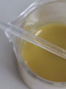
For group 2, 35 % hydrogen peroxide (Opalescence ENDO; Ultradent Products, South Jordan, UT, USA) was applied to the cavities. The occlusal surfaces of the teeth were then covered with plastic wrap. The teeth were stored in a closed container with 100 % humidity at 37 Celsius for 4 days. After the completion of 4-days bleaching process, the teeth were washed with air-water spray for 60 seconds. 10 % vitamin C suspension was prepared by dissolving 1 vitamin C tablet which contained 500 mg of ascorbic acid and sodium ascorbate (Hicee, Takeda Pharmaceutical, Takeda, Tokyo, Japan) into 5 ml 37 degree Celsius distilled water (Figure 2). The cavities were subsequently irrigated with 10 % vitamin C suspension for 10 minutes with a syringe and then agitated gently with micro brush. Following that, the cavities were irrigated with 37 degree Celsius distilled water for 30 seconds, air dried, then restored with resin composite following the previous protocol mentioned in group 1.

The sample preparation for group 3 followed the same bleaching protocol as mentioned earlier. The cavities were restored with resin composite immediately following the same protocol as groups 1 and 2.
After the restoration in all groups were completed, the teeth were stored for 24 hours in a closed container with 100 % humidity at 37 degrees Celsius, and then serially sectioned into 1-mm thick vertical slabs with the water-cooled diamond blade. The specimens were then rotated at 90 degrees and sectioned again to obtain a rectangular dentin-composite stick with approximately 1 mm2 cross sectional area. Four dentin-composite sticks were obtained from each tooth. The specimens were then attached to the micro tensile tester (EZ-S, Shimadzu, Japan) by using cyanoacrylate glue (Cyanoacrylate glue: model repair ll Blue, Densply, Japan). The specimens were then subjected to tensile force at crosshead speed of 1mm/min. The micro tensile bond strength values were expressed in megapascal (MPa) based on the following calculation: force magnitude at which debonded occurred (N) divided by the dentin-composite specimen interface area (mm2).
The mode of failures of the specimens were inspected by the use of stereomicroscope at 40x magnification. Mode of failures were classified into the 4 categories:
- Adhesive failure: the fracture occurred at the interface between dentin and adhesive for more than 70 %.
- Cohesive in dentin: the fracture occurred in the dentin for more than 70 %.
- Cohesive in resin composite: the fracture occurred in the resin composite more than 70 %.
- Mixed failure: a mixed fracture within each structure more than 30 %.
Results
Micro tensile bond strength
The micro tensile bond strength from each group are presented in table 1. Statistical analysis was performed with SPSS Statistics v21.0 (IBM, Armonk, NY) using One-way ANOVA test revealed that group1 (No bleaching) showed significantly highest micro tensile bond strength, followed by group 2, and group 3 respectively. Though bleaching decreased the micro tensile bond strength, 10 % Vitamin C suspension treatment significantly improved bond strength. However, the bond strength was not equal to non-bleached dentin.
Table1
|
Groups (N=40/group) |
Mean ± SD (MPa) |
|---|---|
|
Group 1 : No bleaching Group 2 : Bleaching + Vitamin C Group 3 : Bleaching no vitamin C |
36.417 ± 3.943 a 31.756 ± 4.961b 24.468 ± 5.189 c |
*Value with difference letter are significantly different from each other (p < 0.05)
Mode of failure
Mode of failure from each group are presented in figure 3. The most common failure mode was adhesives failure followed by cohesive failures in dentin.

Discussion
The result of this study revealed that non-vital bleaching affected the micro tensile bond strength of dentin and resin composite by decreasing the bond strength. Titley et al, evaluated the scanning electron microscope (SEM) images of interfaces between resin and bleached enamel and observed fragmented and poorly refined resin tags that penetrated to a lesser depth when compared with unbleached controls. Also resin-bleached enamel interface was granular and porous with a bubbled appearance due to the entrapment of peroxide ions into the bleached enamel.6 Some researchers found that changes in organic substance, loss of calcium, and a decrease in microhardness of tooth structure may contribute this effect.7-9 These could possibly explain the lowest micro tensile bond strength obtained from group 3.
If reduction of bond strength in bleached tooth is a result of the oxidant action of the bleaching agent, it could theoretically be reversed by the application of an antioxidant. In this study the use of 10 % vitamin C suspension could reverse the negative effects of bleaching on bond strength of dentin and resin composite. This may due to the antioxidant capacity of vitamin C. This is consistent with previous studies that have used antioxidant to enhance the bond strength of resin composite and tooth structure. Turkun and Kaya demonstrated that the use of an antioxidant agent could provide normal values in shear bond tests. After the tooth was bleached by carbamide peroxide.16 Kimyai and Valizadeh evaluated the use of hydrogel and sodium ascorbate solution and found no difference of the efficacy between hydrogel and solution formulation.12 This study prepared the 10 % vitamin C suspension by dissolving a vitamin C tablet that contain 500 mg of ascorbic acid and sodium ascorbate with 5 ml distilled water. Most of studies had used 10 % sodium ascorbate according to a study by Dabas which recommended the concentration of 10 % sodium ascorbate, since there was no significant difference in the bond strength of bleached tooth structure and resin composite after using of 10 % and 35 % sodium ascorbate.4
However, micro tensile bond strength of the group treated with vitamin C suspension was still lower than non-bleached group. This is consistent with a study of Hansen and colleagues who found that micro tensile bond strength of non vital bleached dentin treated with 35 % sodium ascorbate was lower than non bleached dentin17, but other researchers were contradictory. Turkun and colleagues which found that the shear bond strength of the non-bleached enamel was higher than bleached enamel treated with 10 % sodium ascorbate.18 Kunt demonstrated the use of an anti-oxidant agent or immersion in saliva could provide normal values in shear tests when compare bleached tooth to non-bleached tooth.19 The cause of the difference in each studies may come from several factors including the quality of the tooth structure, the specimens’ storage protocol, adhesive technique, and bond strength testing method. Lai and colleagues had also recommended to soak the specimen treated with sodium ascorbate in distilled water for ten minutes before testing bond strength to dissolve the crystal formed by sodium ascorbate as observed through the SEM.13 This study rinsed the specimen with distilled water for 30 seconds using syringe. This may be another reason the bond strength of the Vitamin C group lower than non-bleached group. In this study, 10 % vitamin C suspension prepared from Vitamin C tablets was also used. The concentration of antioxidant and purity of the substance may affect the antioxidant properties of the suspension.
Most of the specimen of three groups demonstrated adhesive failure. This may due to the benefit of micro tensile testing that the stress is concentrated at the interface of the specimen and the force is perpendicular to the interface. The fracture usually begins at the weakest point.20,21 This result correlated with Lai et al, which also found that most of the failures were adhesive failures.13 However, group 1 demonstrated more cohesive failures than the other groups. This may be inferred that group 1 demonstrated the highest bond strength, which may more than the cohesive strength of dentin and resin composite, resulting in cohesive fracture within substrate. The SEM would be more beneficial to analyze the structures of the bond interfaces, such as the change in tooth structures or the character of the hybrid layer. Viera and colleagues found that fracture occurred within the hybrid layer and adhesive in tooth specimen bleached with hydrogen peroxide. The study also found the rhomboidal crystals in group treated sodium ascorbate which may affect the bonds strength to adhesive.5
Sodium ascorbate is an antioxidant with low toxicity and commonly use in food industry. However, sodium ascorbate is not commercially available, unlike vitamin C. Some researchers were concerned about the acidity of the ascorbic acid in vitamin C tablet which may cause the demineralization of tooth structures.14-15 The pH of sodium ascorbate is around 7, while ascorbic acid is around 4.14,22 However nowadays, there are vitamin C tablets which the manufactures claimed to be less acidic since a salt form of ascorbic acid was used. In this study the pH of vitamin C suspension prepared by vitamin C tablet was 6.27 which may not be detrimental to the mineralized tooth structures.
The limitations of this study include limited sample of vitamin C tablet used, since only 1 brand was tested. This may not be sufficient to represent all commercially available vitamin C tablets. More studies could be conducted to discuss the effects of vitamin C concentration and the application time of vitamin C suspension to the bleached dentin for clinical benefits in the near future.
Conclusion
Within the limitations of this study, it could be concluded that non-vital tooth bleaching affected the micro tensile bond strength between dentin and resin composite. The bond strength was reduced significantly compared to non-bleached dentin. The use of 10 % vitamin C suspension prepared from vitamin C tablets significantly improved the bond strength of bleached dentin, though the bond strength was lower than the non-bleached dentin. Vitamin C suspension may serve as an alternative of sodium ascorbate to treat non-vitally bleached dentin prior to restoring with resin composite immediately