บทความ
Evaluation of the Posterior Superior Alveolar Artery and Related Factors Using Cone Beam Computed Tomography Images
Abstract
The aim of this study was to assess the prevalence, location, diameter, and distance of the posterior superior alveolar artery (PSAA) from the alveolar crest; and its association with sex, age, and dental status using cone beam computed tomography (CBCT). Retrospectively reviewed CBCT images of maxillary sinuses from 370 sinuses in 185 patients at the Faculty of Dentistry, Chulalongkorn University were evaluated. The prevalence, location, diameter, and the distance between the alveolar crest and PSAA were evaluated. The prevalence, location, diameter, and distance from the alveolar crest were compared according to sex, age, and dental status. The result revealed that the PSAA was detected in 32.16 % of sinuses and most (52.85 %) had an intraosseous location. The diameter was mostly <1 mm (80.31 %). Males had a significantly larger diameter than females (p < 0.05). Most PSAAs (65.28 %) were >15 mm from the alveolar crest. The distance from the alveolar crest was highest at the first premolar and lowest at the first molar. Most detectable PSAAs were <1 mm in diameter and ≥15 mm from the alveolar crest, indicating a low chance of severe bleeding during sinus augmentation. However, a preoperative CBCT is necessary to avoid complications and design an accurate treatment plan.
Correspondence to:
Pornchai Jansisyanont, Department of Oral and Maxillofacial Surgery, Faculty of Dentistry, Chulalongkorn University, 34 Henri-Dunant Road, Wangmai, Pathumwan, Bangkok, 10330, Thailand. Tel: 02-2188581, Email: Jpornchai@hotmail.com
The presence of the maxillary sinus above the surgical site makes placing implants complex. After losing the upper posterior teeth, bone resorption causes an expansion of the maxillary sinus, resulting in inadequate bone height to support a dental implant.1 Consequently, a sinus augmentation or sinus lift is required to elevate the sinus floor to increase the vertical height for successful implant placement.2 The posterior superior alveolar artery (PSAA) runs through the lateral wall of the maxillary sinus and supplies the posterior maxillary teeth, the maxillary sinus, and the Schneiderian membrane.3 Sinus augmentation may damage the artery during bony window preparation and can lead to hemorrhage. Bleeding will obscure the vision of the surgical field and enhance the chance of maxillary sinus membrane perforation.4 The risk of bleeding is directly related to the size of the artery. The greater the size of the artery, the greater the risk of bleeding during surgical manipulation.5 The risk for intra-surgical bleeding is higher for vessels larger than 2 mm.6 Thus, understanding its diameter, location, and distance from the alveolar crest is essential to avoid damaging the artery, decrease complications, and increase the success rate of sinus surgery.
There are many imaging modalities to evaluate the maxillary sinus, such as a panoramic radiograph, Water's view, computed tomography (CT), magnetic resonance imaging (MRI), and cone-beam computed tomography (CBCT). CBCT presents clear images of highly contrasted structures and is useful for evaluating bone. In addition, using CBCT offers more advantages compared with CT. This technique has advantages of its cost-effectiveness, shorter scan time, higher resolution, excellent spatial resolution, easier operation, and lower radiation dose compared with CT.7-9 Moreover, CBCT can detect the PSAA more often compared with CT.10 Therefore, CBCT is a diagnostic tool that is appropriate for evaluating the structure and assistance in the treatment plan or risk assessment.
Several studies found that the PSAA prevalence ranged from 48.6–89.3%.5,11-23 However, other studies reported different results on the position and diameter of the artery.12-15,18-22,24 Moreover, previous studies that investigated the relation of sex, age, and dental status had disparate results.5,13-15,17,21,22,24,25 Therefore, this study assessed the prevalence, location, diameter, distance of the posterior superior alveolar artery from the alveolar crest; and its relation to sex, age, and dental status. The null hypothesis was that there were no significant differences in the prevalence, location, diameter, and distance of the posterior superior alveolar artery from the alveolar crest based on sex, age, and dental status.
Material and Methods
Study population
CBCT images of patients from May 2017 to May 2019 at the Department of Radiology, Faculty of Dentistry, Chulalongkorn University were retrospectively reviewed. The images were acquired as part of patient care and not for research. The selection criteria were CBCT images that included the maxillary sinus and the patient’s age was ≥ 20 years old. Pathology that affected the measurement and poor-quality CBCT images were excluded. Maxillary sinuses that had been grafted or received implants were also excluded. The study protocol was approved by the Human Research Ethics Committee of the Faculty of Dentistry, Chulalongkorn University, Bangkok, Thailand. (HREC-DCU 2019-058)
Image acquisition
CBCT examination were performed using 3D Accuitomo 170 (J Morita MFG. CORP., Kyoto, Japan) with 80-90 kVp, 5-10 mA, and 17.5 sec exposure and a field of view ≥ 10 x 10 cm. The images were reconstructed with a voxel size of 0.25 mm. Measurements were made using the digital ruler included in the i-Dixel software (version 2.1.7.3 J morita MFG.CORP 2010, Kyoto, Japan). One examiner reviewed the CBCT scans under the close supervision of an experienced radiologist and an oral and maxillofacial surgeon.
Measurements
The sinuses on both sides were assessed. The patients’ demographic data were recorded; sex, age (20-29, 30-39, 40-49, 50-59, and ≥ 60 years), tooth area (first premolar, second premolar, first molar, and second molar) and dental status (edentulous, partially edentulous, and fully dentate).
The CBCT images were adjusted to make the occlusal plane parallel to the floor in coronal, axial and sagittal plane. The 4 reference points measured were the center of each tooth (the first premolar, second premolar, first molar, and second molar). To locate the reference points in the edentulous area, the length of this area was divided by the number of missing teeth and the center of this distance was measured.
The following parameters were assessed at 4 reference points:
The prevalence of the PSAA was examined in the coronal sections and multiplanar reconstruction was used to confirm that the regions contained an artery.
The location of the PSAA was divided into 3 categories as shown in Figure 1: (1) Intra-sinus or below the sinus membrane; (2) Intraosseous; and (3) Superficial or at the outer cortex of the lateral sinus wall.15


The diameter of the PSAA was measured in millimeters (mm) and categorized into 3 groups: (1) Less than 1 mm; (2) 1-2 mm; and (3) More than 2 mm.19
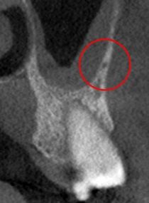
The distance between the most coronal point of the alveolar crest and the lower border of the PSAA was measured vertically in millimeters (mm) and demonstrated in Figure 2. The distance was divided into 2 groups: (1) Less than or equal to 15 mm; and (2) More than 15 mm.20

A = Horizontal line of the lower border of the artery.
B = Horizontal line of the most coronal point of the alveolar crest.
C = The distance of the artery from the alveolar crest.
Statistical analysis
The data were analyzed using statistical software (SPSS version 22.0, IBM, NY, USA). Data were recorded as the frequency (%) and mean ± standard deviation (SD). The detection and location of the arteries were compared according to sex, age range, and dental status using the Chi-square test. The independent t-test, Mann Whitney U test, and Kruskal Wallis H test were used to compare the diameter between sex, age, and dental status. The distance of the artery from the alveolar crest was compared regarding sex, age, and dental status using the independent t-test, one-way ANOVA, and Kruskal Wallis H test. A p-value of less than 0.05 was considered significant.
The intraobserver reliability was determined by reassessing 40 sinuses after more than 2 weeks. The Kappa coefficient was used to calculate the reliability coefficients of the prevalence and location of the artery. Diameter and distance of the artery were calculated by using the Intraclass correlation coefficient.
Results
CBCT images from 185 patients (370 sinuses) were evaluated in this study, there were no significant differences between side, thus the data from both sides were combined. The patients comprised 23.24 % males and 76.76 % females. Their mean age was 48.66 ± 16.6, ranging from 20-87 years. The majority of the patients were fully dentate (61.62 %) and ≥ 60 years old comprised the largest percentage (31.89 %) (Table 1). 149 sinuses (40.27 %) on the CBCT images presented the maxillary sinus variation that did not extend anteriorly to the first premolar area. The intraobserver reliability coefficients ranged from 0.865-1.
Table 1 Sex, age, and dental status distribution of the study population|
Sex |
Na |
% |
Age group |
Na |
% |
Dental status |
Nb |
% |
|
Male |
43 |
23.24 |
20-29 |
39 |
21.08 |
Edentulous |
15 |
4.06 |
|
Female |
142 |
76.76 |
30-39 |
18 |
9.73 |
Partially edentulous |
127 |
34.32 |
|
40-49 |
28 |
15.14 |
Fully dentate |
228 |
61.62 |
|||
|
50-59 |
41 |
22.16 |
||||||
|
age; 60 |
59 |
31.89 |
||||||
|
Number of samples Number of sinuses |
||||||||
A PSAA was detected in 32.16 % of the images. The distribution of the PSAAs at different tooth areas are summarized in Table 2. There was no significant relationship between PSAA prevalence and sex, age, or dental status. Although the prevalence in females was higher compared with males, the difference was only significant in the first molar area (p = 0.025) (Table 3). A PSAA was detected in dentate areas more frequently compared with edentulous areas, however, the difference was only significant in the first molar area (p = 0.015) (Table 3).
Table 2 The prevalence and location of the posterior superior alveolar artery at each tooth position|
Tooth area |
Number of sinuses |
Detection |
Location |
||
|
Intra-sinus |
Intraosseous |
Superficial |
|||
|
First premolar |
221 |
4 (1.81 %) |
0 |
4 (100 %) |
0 |
|
Second premolar |
370 |
33 (8.92 %) |
7 (21.21 %) |
25 (75.76 %) |
1 (3.03 %) |
|
First molar |
370 |
54 (14.59 %) |
33 (61.11 %) |
21 (38.89 %) |
0 |
|
Second molar |
370 |
102 (27.57 %) |
49 (48.04 %) |
52 (50.98 %) |
1 (0.98 %) |
|
Total |
370 |
119 (32.16 %) |
89 (46.11 %) |
102 (52.85 %) |
2 (1.04 %) |
|
Tooth area |
Sex |
p-value a |
Dental status |
p-valuea |
||
|
Male |
Female |
Edentulous area |
Dentate area |
|||
|
First premolar |
4 (1.81 %) |
0 |
0.003* |
2 (0.90 %) |
2 (0.90 %) |
0.113 |
|
Second premolar |
12 (3.24 %) |
21 (5.68 %) |
0.062 |
7 (1.89 %) |
26 (7.03 %) |
0.333 |
|
First molar |
19 (5.14 %) |
35 (9.46 %) |
0.025* |
19 (5.14 %) |
35 (9.46 %) |
0.015* |
|
Second molar |
28 (7.57 %) |
74 (20 %) |
0.237 |
22 (5.95 %) |
80 (21.62 %) |
0.286 |
|
a Chi-square tests * Significant (p < 0.05) |
||||||
The locations of the PSAA in different areas are shown in Table 2. In the second premolar area, most PSAA (75.76 %) was located intraosseous, while majority of PSAA (61.11 %) located intra-sinus for the first molar area. The most frequent location was intraosseous (52.85 %), followed by intra-sinus (46.11 %), and superficial (1.04 %). The location was not significantly associated with sex, age, and dental status.
The mean diameter of the PSAA was 0.84 ± 0.2 mm (range 0.38-1.82 mm). The diameter of the PSAA at different areas is presented in Table 4. The diameter of the PSAA was less than 1 mm in most patients (80.31 %) and 19.69 % demonstrated diameters between 1 and 2 mm. The association between the diameter and age and dental status were not significant. The diameter was significantly different between sexes, except in the first premolar and the first molar area. Males had a significantly a greater mean PSAA diameter compared with females (Table 4).
Table 4 The number of the posterior superior alveolar artery according to diameter and the mean diameter according to sex at each tooth position.|
Tooth area |
Diameter of PSAA |
||||||
|
<1 mm |
1-2 mm |
>2mm |
Mean ± SD |
Male |
Female |
p-value a |
|
|
First premolar |
3 (75%) |
1 (25%) |
0 |
1.06 ± 0.51 |
1.06 ± 0.51 |
- |
-b |
|
Second premolar |
28 (84.85%) |
5 (15.15%) |
0 |
0.86 ± 0.26 |
1.01 ± 0.35 |
0.78 ± 0.14 |
0.041* |
|
First molar |
41 (75.93%) |
13 (24.07%) |
0 |
0.90 ± 0.20 |
0.94 ± 0.23 |
0.88 ± 0.18 |
0.82 |
|
Second molar |
83 (81.37%) |
19 (18.63%) |
0 |
0.84 ± 0.23 |
0.94 ± 0.29 |
0.80 ± 0.20 |
0.017* |
|
Total |
155 (80.31%) |
38 (19.69%) |
0 |
0.84 ± 0.20 |
0.93 ± 0.24 |
0.81 ± 0.17 |
0.013* |
|
PSAA, Posterior superior alveolar artery; SD, Standard deviation a Mann Whitney U test b cannot calculate because only male demonstrated arteries at the first premolar area * Significant (p < 0.05)
|
|||||||
The mean distance of the PSAA from the alveolar crest was 16.75 ± 3.7 mm. The mean distance at 4 positions was determined (Table 5). Figure 3 displays the course of the PSAA, with its highest distance from the alveolar crest at the first premolar that decreased to the lowest distance at the first molar area. The distance did not significantly associate with sex, age, and dental status. Although the dentate areas had a higher distance compared with edentulous areas, the distance was only significant at the first molar area (p = 0.000) (Table 5). Most arteries in this study demonstrated a distance to the alveolar crest more than 15 mm (65.28 %) (Fig. 4) and the majority had diameters less than 1 mm (51.81 %).
Table 5 The mean distance between the alveolar crest and the posterior superior alveolar artery at each tooth position according to dental status|
Tooth area |
Distance (Mean ± SD) |
Dental status |
p-value a |
|
|
Edentulous area (Mean ± SD) |
Dentate area (Mean ± SD) |
|||
|
First premolar |
20.22 ± 7.77 |
13.52 ± 1.03 |
26.92 ± 0.87 |
- |
|
Second premolar |
18.77 ± 4.06 |
16.65 ± 4.83 |
19.34 ± 3.73 |
0.120 |
|
First molar |
15.63 ± 3.69 |
13.29 ± 3.88 |
16.89 ± 2.92 |
0.000* |
|
Second molar |
16.43 ± 3.98 |
16.39 ± 5.13 |
16.44 ± 3.64 |
0.959 |
|
SD, Standard deviation a Independent t-test - cannot calculate due to low sample number at this area * Significant (p < 0.05)
|
||||

Figure 3 The distribution of the mean distance (mm) from the posterior superior alveolar artery to the alveolar crest at 4 tooth positions

Figure 4 The distribution on the prevalence of the posterior superior alveolar artery according to the distance from alveolar crest ≤ 15 mm and >15 mm
Discussion
The present study evaluated the prevalence, location, diameter, and distance of the posterior superior alveolar artery from the alveolar crest based on sex, age, and dental status. The results indicated that there were some significant differences in these parameters among sex, age, and dental status. Based on these results, the null hypothesis was rejected.
In previous studies the PSAA prevalence ranged from 48.6–89.3 %, whereas, the current study found a lower prevalence (32.16%). 5,11-23 The different results may be due to differences in the racial variations and sample sizes among studies. In addition, the lower radiographic detection of the PSAA might be related to their small diameter. Our study used CBCT images with a voxel size of 0.25 mm. Thus, arteries with a diameter lower than 0.25 mm could not be detected. However, studies in cadavers revealed that the PSAA presented in 100 % of the specimens.3,26 This finding indicated that although a PSAA was not found for every patient via CBCT analysis, there was an artery present. Because our study measured at peripheral artery with small diameter and might have been below the sensitivity of the CBCT.22
The prevalence of a PSAA in females was greater compared with males, however, the difference was not significant. In contrast Kim et al.17 and Yalcin & Akyol22 found a significantly higher prevalence in males. This difference may be due to the different ratio of males and females in this study. Other studies demonstrated that older individuals were more likely to have a PSAA,5,22 whereas our study found no relationship between the PSAA prevalence and age. In dentate areas, a PSAA was detected more often compared with edentulous areas, with the detection in the first molar area being significantly higher. This result concurred with Ilguy et al.’s study.14
The location of the PSAA was mostly intraosseous (52.85%), followed by intra-sinus (46.11 %), and superficial (1.04 %), which conformed with the results of prior studies.12-15,20,22,24 However, Lozano-Carrascal et al.18 found that the PSAA was most commonly located below the sinus membrane (53.85 %). Other studies showed a significant relationship between the location of the artery and sex14,21; however, our study did not find this relationship.
In the present study, the most common PSAA diameter (80.31 %) was less than 1 mm. This result concurred with previous studies.14,18,19,24 However, our results were different from other studies that found the most common diameter was between 1 and 2 mm and ranged from 55.8–74.8 %.12,13,20,21 Although, the measurement of PSAA diameter in this study was similar to other studies.12-14,18,20-24 The different diameter might be depending on individual subject. Maridati et al.6 reported that damaging an artery less than 2 mm did not affect the execution of the surgical procedure. We did not find PSAA’s with a diameter above 2 mm. Moreover, most arteries were less than 1 mm. Therefore, these results suggested that this study population would have a low occurrence of severe bleeding during sinus surgery.
The PSAA diameter was significantly different between sexes, except in the first premolar and the first molar area. Males had a larger artery diameter compared with females, which was in accordance with other reports.5,13,21,24 The current study found no relationship between PSAA diameter and age or dental status that matched the findings of Guncu et al.15 and Danesh-Sani et al.13 However, other studies demonstrated that older individuals had a greater artery diameter.5,25
The mean distance from the PSAA to the alveolar crest was 16.75 ± 3.7 mm, which was similar to the findings of Mardinger et al.19 (16.9 ± 4.4 mm), Tehranchi et al.21 (16.7 ± 3.96 mm), Chitsazi et al.12 (16.17 ± 1.63 mm), and Simsek Kaya et al.20 (16.95 ± 1.9 mm). These studies12,19,20 use the same measurement with this study except Tehranchi et al.21 measured with the closest distance to alveolar ridge or did not measure vertically. The present study revealed that dentate areas had a higher distance compared with edentulous areas. This finding was in concordance with Velasco-Torres et al.25, who found that the distance from the alveolar crest reduced when teeth were lost. This might be because tooth loss caused alveolar ridge resorption and decreased the distance between the PSAA and alveolar crest. The sex and age of the subjects did not significantly affect this distance in our study. In contrast, Khojastehpour et al.5 and Tehranchi et al.21 reported that males had a significantly greater distance compared with females. Moreover, our results demonstrated that the course of the PSAA had the highest distance from the alveolar crest at the first premolar and decreased to its most inferior level in the first molar area. These results are similar to Kim et al.17, Jung et al.16 and Aung et al.23 who demonstrated that the PSAA was closer to the alveolar crest in the molar area.
The lateral wall sinus lift technique uses a superior horizontal osteotomy line up to 15 mm from the alveolar crest, which is adequate for exposing the lateral wall of the sinus surgery and placing dental implants.19,27 We found that most PSAAs had a distance to the alveolar crest of more than 15 mm (65.28 %). The PSAA in a distance less than 15 mm was seen in only 5.95 % of images in the first molar area and a lower prevalence in the first premolar and the second premolar area of 0.9 % and 1.62 %, respectively. These PSAAs could present a low occurrence of hemorrhage in sinus surgery. If the osteotomy line is more than 15 mm, the surgeon should concern about potential bleeding during surgery. Therefore, the surgeon should plan the surgery using CBCT to avoid damaging the artery and be prepared to control bleeding if it occurs.
A limitation of this study was that some CBCT images presented the anterior wall of the maxillary sinus that did not extend to the first premolar area (40.27 %). These images might affect the PSAA prevalence in the first premolar area where only 4 arteries were found. These limited samples could not be statistically analyzed. Moreover, the lower number of males compared with females might not have been sufficient to identify a significant difference based on sex. Accordingly, further studies should have equal number of males and females and the maxillary sinus should include all posterior maxillary teeth.
Conclusion
Based on our findings, most of the PSAAs had a diameter less than 1 mm and the distance of the artery from the alveolar crest was more than 15 mm that should make a low chance of severe bleeding during surgery. However, the PSAA course and size is different in each individual. Thus, comprehensive planning using CBCT is necessary prior to surgery to avoid complications during surgery and develop an accurate treatment plan.
Acknowledgments
The authors greatly appreciate the help of Assist. Prof. Dr. Soranun Chantarangsu in statistical analysis and Dr. Kevin Tompkins for English revision.
Nutcha Benjaphalakron1, Pornchai Jansisyanont1, Vannaporn Chuenchompoonut2, Sirichai Kiattavorncharoen3
- Department of Oral and Maxillofacial Surgery, Faculty of Dentistry, Chulalongkorn University, Bangkok, Thailand
- Department of Radiology, Faculty of Dentistry, Chulalongkorn University, Bangkok, Thailand
- Department of Oral and Maxillofacial Surgery, Faculty of Dentistry, Mahidol University, Bangkok, Thailand